Arteries Diagram : Anatomy Of Lower Extremity Leg Artery Supplement Diagram - The pulmonary trunk and arteries of the pulmonary circulation loop provide an exception to this rule — these arteries carry deoxygenated blood from the heart to the lungs to b
Arteries Diagram : Anatomy Of Lower Extremity Leg Artery Supplement Diagram - The pulmonary trunk and arteries of the pulmonary circulation loop provide an exception to this rule — these arteries carry deoxygenated blood from the heart to the lungs to be oxygenated.. Most arteries carry oxygenated blood; Main branches from the aorta include the brachiocephalic artery, left carotid artery, and the left subclavian artery. The incidence of anatomical variations regarding celiac trunk. The transverse cervical artery is the next branch off the thyrocervical trunk. Diagram of the human circulatory system (infographic).
The arteries of the brain; An artery is a blood vessel that takes blood away from the heart to one or more parts of the body. This supplies blood to the knee region. Main branches from the aorta include the brachiocephalic artery, left carotid artery, and the left subclavian artery. The arteries are part of the circulatory system, which is responsible for the delivery of oxygen and nutrients to all cells, as well as t
Artery Diagram High Resolution Stock Photography And Images Alamy from c8.alamy.com It originates from the heart and branches out into smaller arteries which supply blood to the head region (brachiocephalic artery), the heart itself (coronary arteries), and the lower regions of the body. There are two paired arteries which are responsible for the blood supply to the brain; Each artery is a muscular tube lined by smooth tissue and has three layers: Brachiocephalic trunk, left common carotid artery and left subclavian artery. Arteries of the brain and 'circle of willis' diagram. This supplies blood to the knee region. The arteries of the brain; The right coronary artery supplies blood to the right ventricle, the right atrium, and the sa (sinoatrial) and av (atrioventricular) nodes, which regulate the heart rhythm.
The first branch of the thyrocervical trunk is the inferior thyroid artery.
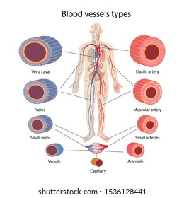
Systemic arteries deliver blood to the rest of the body. This is known as the main pulmonary artery or pulmonary trunk. Circle of willis is indeed a hot neuroanatomy topic! This vessel supplies the posterior prevertebral muscles. The two exceptions are the pulmonary and the umbilical arteries, which carry deoxygenated blood to the organs that oxygenate it. Lungs (pulmonary), and arteries, veins, coronary and portal vessels (systemic). The capillaries connect the two types of blood. The system is responsible for the flow of blood, nutrients. Human anatomy for muscle, reproductive, and skeleton. The arteries of the brain; See the back for a diagram showing the two circulation routes. The arteries of the upper extremity the subclavian artery; Blood carried by arteries is usually highly oxygenated, having just left the lungs on its way to the body's tissues.
Next, we have the blood vessel responsible for carrying deoxygenated blood from the right side of the heart (right ventricle) to the lungs. Lungs (pulmonary), and arteries, veins, coronary and portal vessels (systemic). Brachiocephalic trunk, left common carotid artery and left subclavian artery. The pulmonary trunk and arteries of the pulmonary circulation loop provide an exception to this rule — these arteries carry deoxygenated blood from the heart to the lungs to be oxygenated. It is a central communication that unites the internal carotid and vertebrobasilar systems.
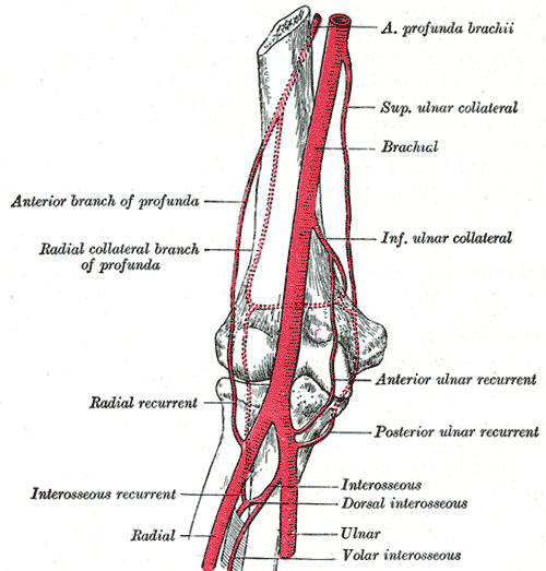
Arteries Of The Upper Limb The Lecturio Medical Online Library from d3uigcfkiiww0g.cloudfront.net There is a point at which the anterior and posterior arterial circuits of the brain unite or anastomose. Well you're in luck, because here they come. The arteries of the upper extremity the subclavian artery; Blood is pumped from the heart in the arteries. 28 vein artery diagram artery and vein diagram arteries and. This area is known as the circle of willis. See the back for a diagram showing the two circulation routes. The two exceptions are the pulmonary and the umbilical arteries, which carry deoxygenated blood to the organs that oxygenate it.
The first branch of the thyrocervical trunk is the inferior thyroid artery.
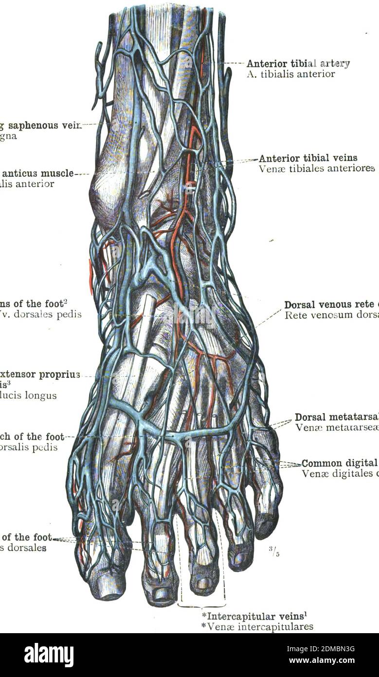
Blood is transported in arteries, veins and capillaries. The right coronary artery divides into smaller branches, including the right posterior descending artery and the acute marginal artery. Several branches of the external iliac artery extend into the abdominal, groin, and pelvic regions, but the bulk of its blood continues onward into the leg, where it becomes known as the femoral artery. Well you're in luck, because here they come. Lungs (pulmonary), and arteries, veins, coronary and portal vessels (systemic). The main pulmonary artery splits into the right and left pulmonary arteries (better seen in the diagram at the end of this post). It is returned to the heart in the veins. See the back for a diagram showing the two circulation routes. The left coronary cusp gives rise to the left main coronary artery which branches to the left anterior similarly, branches from the lcx are labelled obtuse marginal and numbered in order. The arteries of the upper extremity the subclavian artery; The arteries are part of the circulatory system, which is responsible for the delivery of oxygen and nutrients to all cells, as well as t There is a point at which the anterior and posterior arterial circuits of the brain unite or anastomose. The ascending cervical artery arises from the inferior thyroid artery, as it turns medially in the neck.
The aorta is the largest artery in the body that exits the left ventricle of the heart. Amicus illustration of amicus,anatomy,brain,arterial,artery,arteries,supply,cerebral,cavernous the right and left upper and lower limbs create a flow chart showing the major systemic veins through which blood travels… Most arteries carry oxygenated blood; Arteries of the brain and 'circle of willis' diagram. Blood is transported in arteries, veins and capillaries.
Arteries Veins Capillaries Diagram High Res Stock Images Shutterstock from image.shutterstock.com An artery is a blood vessel that takes blood away from the heart to one or more parts of the body. This is known as the main pulmonary artery or pulmonary trunk. The aorta is the main systemic artery and the largest artery of the body. The triangles of the neck; The two exceptions are the pulmonary and the umbilical arteries, which carry deoxygenated blood to the organs that oxygenate it. This supplies blood to the knee region. It is returned to the heart in the veins. It is a central communication that unites the internal carotid and vertebrobasilar systems.
The right coronary artery divides into smaller branches, including the right posterior descending artery and the acute marginal artery.
Circle of willis is indeed a hot neuroanatomy topic! An artery is a blood vessel that takes blood away from the heart to one or more parts of the body. The human body arteries and veins diagram can be major systemic arteries. The arteries of the head and neck. 5.5 / 10 ( 2 votes ) blood circulation principal veins and arteries diagram. Each artery is a muscular tube lined by smooth tissue and has three layers: The left coronary cusp gives rise to the left main coronary artery which branches to the left anterior similarly, branches from the lcx are labelled obtuse marginal and numbered in order. The vertebral arteries, and the internal carotid arteries. The incidence of anatomical variations regarding celiac trunk. The brachiocephalic trunk gives rise to the right common carotid and right subclavian arteries. The media, a layer of muscle that lets arteries. Head and neck arteries (diagram) blood supply for the head and neck comes from the branches of the aortic arch: There are two paired arteries which are responsible for the blood supply to the brain;


Comments
Post a Comment